PET Center
Positron emission tomography (PET)是一种专门的放射学程序,它使用分子成像来跟踪和追踪正常和异常情况. PET is the most advanced nuclear imaging technique, 允许高分辨率成像和绝对定量的生物机制. PET成像也用于跟踪患者接受治疗的进展. While PET is most commonly used in the fields of neurology, oncology and cardiology, applications in other fields are currently being studied.
On This Page
Our Mission
Our mission is to lead in discovery, PET药物的教学和应用,以及公平分配和改进以人为本的护理方法.
About PET
约翰霍普金斯大学PET放射性示踪剂中心自1983年安装第一台回旋加速器以来一直在持续运行. 它是发现新型放射性示踪剂的主要中心之一, 实施在其他地方发现的有前途的新放射性示踪剂,并管理我们社区的患者.
PET是一种分子成像技术,从小动物临床前研究中无创地测量生物化学
s to human beings. 这项技术的工作原理是通过检测正电子的湮灭,在给予正电子发射的放射性示踪剂后,正电子发生在一个特定的受体上, transporter, enzyme or antigen. 当湮灭光子巧合地到达扫描仪中放置在推荐十大正规网赌平台周围的相反探测器时,就会被检测到. 从检测到的光子离开身体时的位置重建图像,从而产生在疾病部位和整个身体中发生的生化活动的图像. 自20世纪80年代以来,PET扫描在临床研究和患者管理中都是一种有价值的技术. 然而,在过去的20年里,医疗保险中心 & 医疗补助服务(CMS)已经报销了临床PET研究, making them widely available in the US.
Rarely are such advanced PET imaging studies performed alone. Currently, PET与提供解剖信息的计算机断层扫描(CT)一起进行,融合的分子和解剖(CT)图像一起解释为PET/CT研究. 这些研究大多利用葡萄糖的正电子发射形式, known as fluorodeoxyglucose (FDG), to check for tumor metabolism. 但我们在临床上进行了许多其他这样的研究,以指导疾病诊断和监测对治疗的反应.
Patient Care
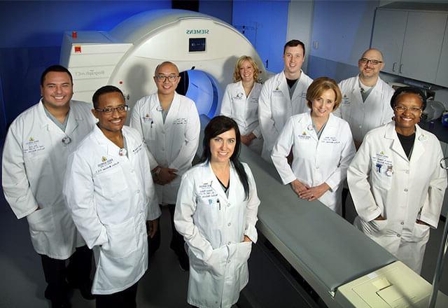
我们的PET中心团队每年进行超过4,300例手术. 我们训练有素的放射科医生管理和解释每一次PET扫描. These advanced, 高分辨率图像使我们的团队和临床合作伙伴能够了解体内器官系统的结构和功能. 我们最先进的设备和技术是我们坚定不移的承诺,提供卓越的患者护理和对细节的关注.
PET scans are often used to help diagnose cancer,检测癌症向身体其他部位的扩散,或测量药物的有效性 cancer treatment. 其他用途包括诊断大脑和心脏疾病,如 Alzheimer's disease, Parkinson's disease, Huntington's disease, epilepsy, stroke, inflammatory heart disease or coronary artery disease.
The Next Generation: PET/CT
最近的技术进步使PET扫描仪与x射线CT结合在一个相机系统中. 这些混合系统通过单一的无创成像研究获得最大的结构和生物信息, combining the power of PET and CT. 我们的扫描有助于诊断、治疗和评估各种状况和疾病
Test Results Online
患者可以在扫描后三天内获得检测结果 MyChart. MyChart是一个安全的网站,为您提供有关约翰霍普金斯医疗保健的最新医疗信息,并将您与您的医疗保健团队联系起来. MyChart is available in English and Spanish. To sign up for MyChart, 您将在预约期间收到唯一的激活码,以创建新的MyChart用户ID和密码.
PET Center Special Services
Below are some clinical scans performed at Johns Hopkins. 根据您接受护理的地点,有些扫描可能不可用.
- PET/CT FDG Oncologic Imaging, 利用代谢生物标志物18F-FDG来帮助诊断癌症并检测癌症向其他器官的扩散, as well measure the effectiveness of anti-cancer therapy.
- PET/CT Sodium Fluoride Imaging, using the F18-NaF biomarker, a highly sensitive bone-seeking PET tracer, to detect skeletal abnormalities.
- PET/CT FDG Brain Imaging, 用于评估认知障碍,痴呆和癫痫.
- PET/CT AMYLOID Imaging, 使用F18- florbetaben和F18- Florbetapir等生物标志物来评估认知障碍患者的淀粉样斑块密度, Alzheimer’s disease and other causes of cognitive decline.
- PET/CT Myocardial Viability Imaging, 18F-FDG评估晚期冠状动脉疾病和左心室功能障碍患者的心肌活力.
- PET/CT Myocardial Perfusion Imaging, 使用N-13氨提供了高灵敏度的冠状动脉闭塞性疾病检测, evaluation of the effectiveness of therapeutic interventions, and evaluation of quantitative myocardial flow reserve.
- PET/CT Inflammation and Infection Imaging, using F18-FDG for detection of sarcoidosis, large-vessel vasculitis, implant related infections, and fever of unknown origin.
- PET/CT Ga68 Dotatate Imaging 生长抑素受体对神经内分泌肿瘤的高敏感检测及治疗随访.
- PET/CT Pylarify PSMA for Prostate Imaging 用于根据血清PSA升高怀疑有转移或复发的患者
- PET/CT Ga68 PSMA for Prostate Imaging similar to Pylarify. 用于治疗评估和将使用Pluvicto治疗的患者.
Rates
For latest rates and services, please download the PDF.
Locations
PET/CT scans are available at The Johns Hopkins Hospital, Johns Hopkins Medical Imaging in Bethesda and Green Spring Station, and Johns Hopkins Bayview Medical Center. Request an appointment by calling 443-997-7237.
Education
Our division offers a 约翰霍普金斯大学医学院的临床PET/CT奖学金项目. 为期一年的奖学金项目提供正电子发射断层扫描/计算机断层扫描(PET/CT)的临床和研究方面的专业培训。. 成立于1999年的acgme认证奖学金直接与 nuclear medicine residency program.
Contact Information
For more information, please contact:
Corina Voicu CNMT RT(CT)
PET Imaging Manager
410-955-2905
Leadership
Lilja Solnes, M.D., M.B.A.
Division Chief, Nuclear Medicine and Molecular Imaging
Martin Pomper. M.D., Ph.D.
Research Chief, Nuclear Medicine and Molecular Imaging
Sridhar Nimmagadda, Ph.D.
Director, PET Center
Robert F. Dannals, Ph.D.
Director, PET Chemistry